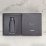
This means that your body is using fatty acids to make energy for the brain. Ketone levels can tell you if your body is in ketosis. The Lumen device does not measure ketones at all instead it is an alternative measurement to ketones, measuring CO2 and this reading can determine whether your body is burning fat or carbs. Lumen will tell you if your body is using carbs or fats as a source of energy. Figure used with permission from the Esophageal Center at Northwestern.A ketone meter, like this one, take a blood sample to measure blood ketone levels – another metabolic tracker. In addition, the contractile activity in the body of the esophagus is erratic with both antegrade and retrograde contractions suggesting a motility disorder.

The concomitant FLIP topography displays the narrowed zone at the EGJ that appears to remain relatively closed through most of the study and never opens to a level greater than 12.5 mm. At time point 4 with a 60 ml filling volume, the diameter is 12.5 mm and the pressure is 42 mmHg with a distensibility index of 2.9 which is right at the cut-off value of 2.8 mm 2/mmHg for achalasia. At time point 3, the EGJ initially opens at an intra-balloon pressure of 24 mmHg and the distensibility index is 1.0 mm 2/mmHg. As the volume distention occurs with filling from 20 ml to 60 ml, the EGJ is visualized on the 3-D images as a narrowing and low diameter. In the achalasia patient (2B), time point 1 once again represents baseline with 5 ml filling. Time point 5 represents a period of quiescence at 60 ml with balloon diameters reaching the limits of the bag at 22mm. Time points 2–4 represent propagation of a single antegrade contraction within the sequence of RACs where one can see the narrowing associated with contraction move distally within the 3-D image and the propagating contraction on FLIP topography noted by the red color similar to peristalsis noted on high-resolution manometry. Time point 1 illustrates the baseline with minimal pressure and balloon filling. In the asymptomatic control (2A), real-time 3-D images are depicted to show various points of interest using shape to highlight narrowing. Diameter data from each impedance planimerty channel is scaled from 5 to 30 mm and is interpolated and color coded on a hot/cold scale (small diameters are red/large diameters are blue) to generate FLIP topography akin to what is visualized during high-resolution manometry. The pressure (red) recordings are depicted in the upper panel. All rights reserved.Įxamples of data acquisition and analysis in an asymptomatic control (A) and an achalasia patient (B). Best Practice Advice 5: FLIP should not be used to diagnose eosinophilic esophagitis but may have a role in severity assessment and therapeutic monitoring.Īchalasia Eosinophilic Esophagitis Functional Lumen Imaging Probe Gastroesophageal Reflux Disease Impedance Planimetry.Ĭopyright © 2017 AGA Institute.

Best Practice Advice 4: Clinicians should not utilize FLIP in routine diagnostic assessments of gastroesophageal reflux disease. Best Practice Advice 3: Utilization should follow distinct protocols and analysis paradigms based on the disease state of interest. Best Practice Advice 2: FLIP assessment is a complementary tool to assess esophagogastric junction opening dynamics and the stiffness of the esophageal wall. Best Practice Advice 1: Clinicians should not make a diagnosis or treatment decision based on functional lumen imaging probe (FLIP) assessment alone.
#Lumen reviews update
This clinical practice update describes the technique and reviews potential indications in achalasia, eosinophilic esophagitis, and gastroesophgeal reflux disease. However, data are accumulating that are providing guidance regarding emerging applications in the evaluation and management of foregut disorders. This is primarily because of a paucity of data supporting its utility in general practice and a lack of standardized protocols and data analysis methodology. Although it has been commercially available since 2009 as the endolumenal functional lumen imaging probe (EndoFLIP), the functional luminal imaging probe has had limited penetrance into clinical settings outside of specialized centers. Additionally, this tool is also approved to guide therapy during bariatric procedures and specialized esophageal surgery. The functional luminal imaging probe is a Food and Drug Administration-approved measurement tool used to measure simultaneous pressure and diameter to guide management of various upper gastrointestinal disorders.


 0 kommentar(er)
